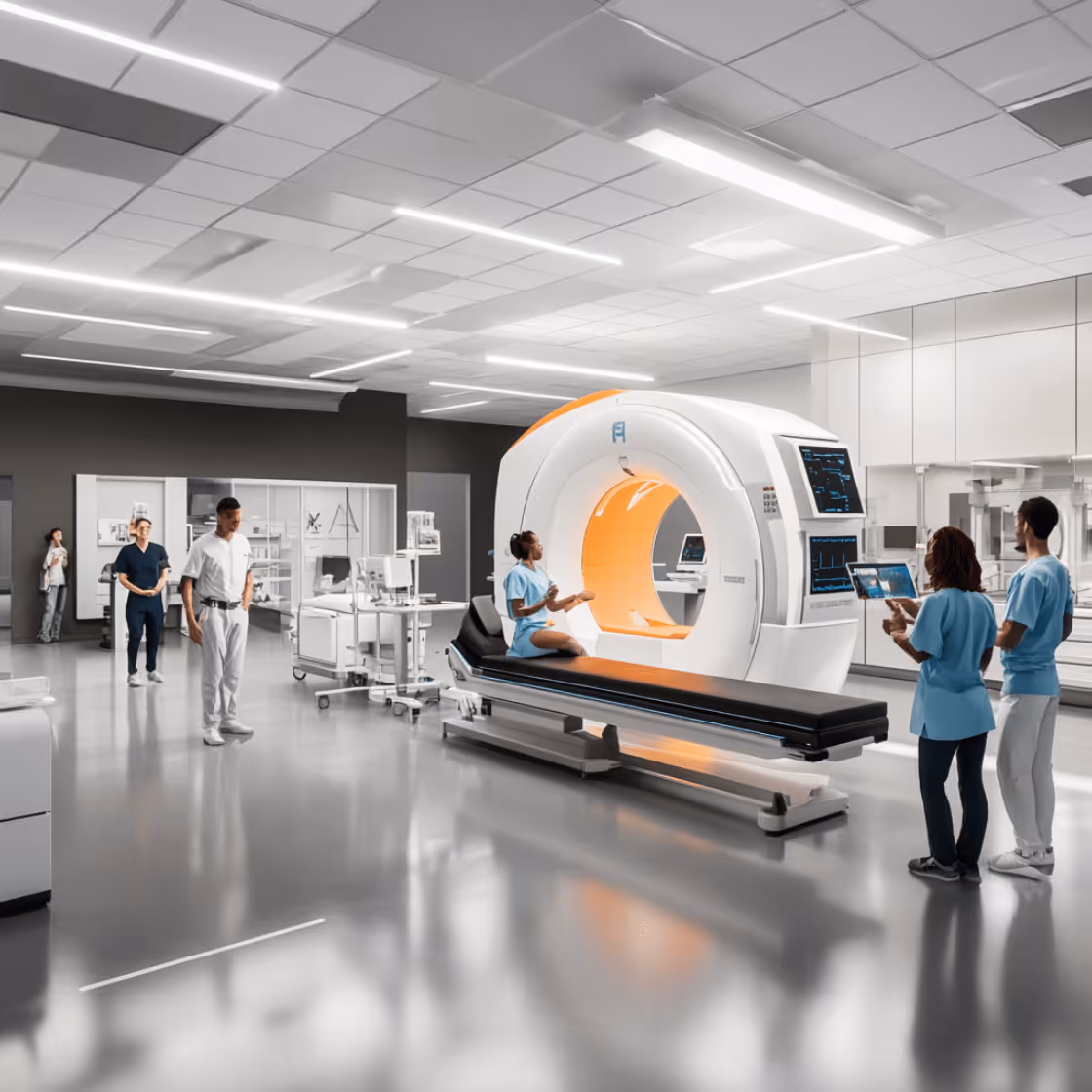
Breast tomosynthesis, also known as 3D mammography or digital breast tomosynthesis (DBT), is an advanced imaging technology used to detect breast cancer with greater accuracy than traditional 2D mammograms. By capturing multiple X-ray images of the breast from different angles and reconstructing them into a three-dimensional view, tomosynthesis allows radiologists to examine breast tissue layer by layer. This technique is especially beneficial for women with dense breast tissue, where overlapping structures in standard mammograms can obscure early signs of cancer.
The procedure follows a structured approach: preparing the patient, positioning and compressing the breast, acquiring multi-angle images, reconstructing the 3D volume, and reviewing and communicating the results. Though it involves slightly more radiation than 2D mammography, the exposure remains within safe limits and is justified by the increase in diagnostic precision.
On average, the cost of breast tomosynthesis ranges from $100 to $250, depending on location, facility type, and insurance coverage. While not universally covered, many insurance providers include 3D mammography in their preventive screening benefits.
More effective at detecting small or hidden tumors, breast tomosynthesis reduces false positives, lowers recall rates, and improves early cancer diagnosis. Compared to standard mammography, it offers a clearer, more comprehensive view of breast anatomy, making it a preferred choice in modern breast cancer screening protocols.
Key Takeaways
- Breast tomosynthesis, or 3D mammography, improves breast cancer detection by providing detailed 3D images, reducing false positives, and enhancing diagnostic accuracy compared to traditional 2D mammograms.
- The procedure for breast tomosynthesis is similar to a standard mammogram but involves taking multiple images from various angles to create a 3D representation, and while it uses slightly more radiation, its benefits outweigh the risks.
- Costs for breast tomosynthesis vary based on location and insurance coverage, averaging between $100 and $250, with many insurance plans partially or fully covering the procedure.
What is breast tomosynthesis?
Breast tomosynthesis, also known as 3D mammography, digital breast tomosynthesis (DBT), or simply tomosynthesis, is an advanced imaging technique used for breast cancer screening and diagnosis. It differs from traditional 2D mammography by capturing multiple low-dose X-ray images of the breast from various angles. These images are then reconstructed into a detailed three-dimensional view of the breast tissue.
This layered imaging approach allows radiologists to examine the breast in fine slices, reducing the visual overlap of tissues that can obscure abnormalities in standard 2D images. As a result, breast tomosynthesis significantly enhances the detection of small tumors, especially in women with dense breast tissue, and decreases the likelihood of false positives. Its clinical accuracy and diagnostic precision have led to widespread adoption in modern breast cancer screening programs.

Is tomosynthesis the same as 3d mammography
Yes, tomosynthesis is the same as 3D mammography. Both terms refer to the same imaging technique that involves taking multiple X-ray images of the breast from different angles and reconstructing them into a three-dimensional image. This method is a significant advancement over conventional mammography, which only captures two-dimensional images.
The primary advantage of 3D mammography over standard mammograms is the ability to see breast tissue more clearly. Creating a layered view of the breast reduces the overlap of tissues that can sometimes obscure abnormalities in 2D images. This leads to more accurate screening mammograms and fewer callbacks for additional mammograms or tests, ultimately providing a more precise diagnostic tool.
How accurate are 3d mammograms?
Research indicates that 3D mammography is highly effective in detecting breast cancer earlier and more accurately than traditional 2D mammography, especially in cases involving dense breast tissue. The technology’s ability to capture three-dimensional images of the breast allows radiologists to identify cancers that might otherwise be missed in standard mammograms.
One of the significant benefits of digital breast tomosynthesis is the reduction in false positives. Traditional mammograms can sometimes lead to false-positive findings, where normal tissue appears suspicious, resulting in unnecessary callbacks for additional testing. 3D mammography has been shown to reduce these false positives, decreasing the anxiety and stress associated with further imaging tests.
The adoption of digital breast tomosynthesis has grown substantially since its approval by the FDA in 2011. Today, nearly half of all mammography machines in the U.S. are equipped with this technology, underscoring its importance in modern breast cancer screening programs. This widespread adoption reflects the trust that breast radiologists and patients place in the enhanced accuracy and reliability of 3D mammograms.

What is the procedure to perform breast tomosynthesis?
Breast tomosynthesis follows a process similar to traditional mammography but incorporates advanced steps to create a three-dimensional image of the breast. The procedure is safe, quick, and typically completed within 15–20 minutes. The process includes the following steps:
- Preparing the patient: Patients may be advised to schedule the exam when breast tenderness is minimal, often in the first half of the menstrual cycle. They are asked to remove any clothing or accessories above the waist and wear a medical gown.
- Positioning and compressing the breast: The patient stands in front of the imaging machine while a technician positions one breast on a flat support plate. Gentle compression is applied to spread out the breast tissue evenly for clearer imaging.
- Acquiring the images: An X-ray tube moves in an arc over the compressed breast, capturing multiple images from different angles in just a few seconds.
- Reconstructing the 3D volume: The images are digitally reconstructed into a layered, three-dimensional view of the breast, allowing radiologists to examine thin slices of tissue individually.
- Reviewing the procedure: A radiologic technologist ensures that image quality is sufficient and that all necessary views have been obtained before ending the exam.
- Communicating the results: A breast radiologist analyzes the images and provides a detailed report. Results are then communicated to the patient or referring physician, typically within a few days.
Below is a breakdown of each step with definitions, execution details, key considerations, and its significance.
1. Preparing the patient
This initial step involves getting the patient ready for the imaging procedure. Patients are typically advised to schedule the exam during the first half of their menstrual cycle to minimize breast tenderness. They are asked to remove clothing from the waist up and wear a gown provided by the facility. Additionally, deodorants, powders, or lotions should not be applied to the underarm or breast area on the day of the exam, as these can interfere with imaging.
Proper preparation enhances patient comfort and helps reduce motion artifacts during imaging. Ensuring that the patient is informed and relaxed also improves cooperation, leading to more accurate results.
2. Positioning and compressing the breast
Once the patient is prepared, a trained technologist positions one breast at a time on a flat support plate. The breast is then gently compressed with a paddle to spread out the tissue evenly. Proper positioning is essential for optimal image quality and complete coverage of the breast tissue.
Compression, though sometimes uncomfortable, is a critical step. It reduces the thickness of the breast, minimizes motion, and helps achieve clearer images by separating overlapping structures. Technologists are trained to balance adequate compression with patient comfort.
3. Acquiring the images
During image acquisition, the X-ray tube moves in a gentle arc over the compressed breast, capturing a series of low-dose images from multiple angles. This typically takes only a few seconds per breast. The patient is asked to remain still and hold their breath briefly during each scan to avoid motion blur.
This step is vital for obtaining high-quality raw data used in the 3D reconstruction. Capturing images from multiple angles reduces the limitations of traditional 2D imaging and allows better visualization of abnormalities that may be hidden by overlapping tissue.
4. Reconstructing the 3D volume
After image acquisition, the system uses specialized software to compile the multiple 2D projections into a three-dimensional dataset. This layered view of the breast enables radiologists to scroll through individual slices, examining the tissue in detail.
Reconstruction transforms raw images into a format that mimics slicing through the breast one layer at a time, reducing anatomical noise. This enhanced view increases the likelihood of identifying early-stage tumors that might not be visible on a single flat image.
5. Reviewing the procedure
Once images are reconstructed, the technologist ensures that all views are complete and meet clinical standards. If necessary, additional images may be taken immediately to address any quality issues or incomplete views. This quality check ensures that the exam is comprehensive before the patient leaves.
Careful review of image quality before finalizing the exam helps prevent the need for repeat visits and improves diagnostic reliability. It is an essential step in maintaining clinical accuracy and patient trust.
6. Communicating the results
The final step involves the interpretation of the 3D images by a breast radiologist. The radiologist evaluates each slice for signs of abnormal tissue, compares findings with prior exams if available, and generates a detailed report. Results are typically shared with the patient or their physician within a few days.
Timely and accurate communication of results is crucial for clinical decision-making and early intervention if abnormalities are detected. It also reduces patient anxiety by ensuring they are informed promptly and clearly about their breast health status.
Advanced breast screening at Fountain Life
At Fountain Life, we combine cutting-edge diagnostics like breast tomosynthesis with whole-body imaging and precision health assessments to catch diseases before symptoms ever appear. Our breast screening services are tailored for women with dense breast tissue or elevated risk factors, offering early, accurate detection and peace of mind. With expert radiologists and AI-supported diagnostics, we go beyond standard screenings to provide truly proactive healthcare.
Does tomosynthesis use more radiation?
Yes, tomosynthesis does use slightly more radiation compared to conventional mammography. However, the increase is minimal and within the safety limits established by regulatory bodies. The additional radiation comes from the multiple X-ray images taken from different angles to create the 3D representation of the breast, which is considered low-dose X-rays.
The benefits of digital breast tomosynthesis, including improved accuracy and reduced false positives, outweigh the slight increase in radiation. The advanced imaging provided by 3D mammography leads to fewer unnecessary biopsies and more precise detection of abnormalities, outweighing the risks associated with the additional radiation.
Is a 3d mammogram less painful?
Yes, a 3D mammogram is often considered less painful than a traditional 2D mammogram. Although both types of mammograms require breast compression to ensure high-quality images, 3D mammography machines are designed with improved comfort features. These may include curved compression paddles that better conform to the natural shape of the breast and reduce pinching. Additionally, 3D imaging is typically completed more quickly, minimizing the time the breast is under compression.
While patients may still experience pressure or mild discomfort, the shorter duration and enhanced equipment design make the process more tolerable for many. Reducing pain and discomfort can also help improve patient compliance with routine screening, which is essential for early breast cancer detection.
What is the cost of breast tomosynthesis?
The cost of breast tomosynthesis typically falls between $100 and $250, with an average cost of approximately $208. This fee generally includes the imaging procedure, use of the 3D mammography equipment, radiologist interpretation of the images, and administrative processing. However, total charges may vary depending on where and how the screening is performed.
Several factors influence the cost of breast tomosynthesis. Geographic location plays a major role—facilities in urban or high-cost-of-living areas often charge more than those in rural settings. The type of medical facility also matters; specialized imaging centers or hospitals may have different pricing structures compared to general clinics.
The level of technology used during the screening can also affect pricing. Centers equipped with the latest 3D mammography systems or artificial intelligence enhancements may charge higher fees. Additionally, the expertise of the radiologist interpreting the results can influence cost, especially if read by a board-certified specialist in breast imaging.
Insurance coverage is another critical factor. Some insurance plans fully cover breast tomosynthesis as part of routine screening, while others may require patients to pay a portion or the full amount out of pocket. To avoid unexpected charges, patients are encouraged to verify pricing and coverage in advance with both the imaging facility and their insurance provider.
Is breast tomosynthesis covered by insurance?
Yes, breast tomosynthesis is often covered by insurance, but the extent of coverage depends on the specific insurance plan and regional regulations. Many private insurance providers and public health programs now include 3D mammography as part of routine breast cancer screening, especially for women over 40 or those at higher risk. In such cases, the procedure may be fully covered with no out-of-pocket costs. However, coverage is not universal—some plans may classify breast tomosynthesis as an enhanced screening and require patients to pay a portion of the cost or meet a deductible first.
Coverage also varies by state. In the United States, several states have enacted legislation requiring insurers to cover 3D mammography at no additional cost to the patient, but this is not yet consistent nationwide. Therefore, patients should confirm their benefits directly with their insurance provider and the imaging facility before scheduling the exam.
If breast tomosynthesis is not covered, patients may choose to appeal the decision or seek advocacy support, especially if the screening is medically necessary due to dense breast tissue or a family history of breast cancer.
Why do you need a breast tomosynthesis?
Breast tomosynthesis is a vital tool for detecting breast cancer, particularly in its early stages. For women with dense breast tissue, traditional 2D mammograms can sometimes miss small abnormalities due to the overlapping of dense tissue. Digital breast tomosynthesis, with its ability to create a 3D image of the breast, provides a clearer and more detailed view, enhancing the detection of early breast cancer.
Moreover, breast tomosynthesis reduces the number of false positives and unnecessary biopsies, leading to fewer call-backs and less anxiety for patients. It is particularly beneficial for women with a strong family history of breast cancer, as it can detect multiple breast tumors that might be missed by conventional mammography. Breast tomosynthesis offers better visualization of whole breast tissue, playing a crucial role in effective cancer screening and detection.

Get personalized, early detection with Fountain Life
If you're looking for a proactive approach to breast health, Fountain Life provides access to advanced screening tools, including 3D mammography, as part of our comprehensive early detection programs. Our data-driven care model identifies risk factors long before they become problems, empowering you to take control of your health through timely, personalized interventions.
What is the difference between a 3D mammogram and a 2D mammogram?
The primary difference between a 3D mammogram and a 2D mammogram lies in how the breast images are captured and analyzed. A 2D mammogram takes two flat X-ray images, one from the top and one from the side, compressing all breast tissue into a single layer. This can sometimes make it difficult to distinguish overlapping structures and may obscure small tumors, particularly in women with dense breast tissue.
In contrast, a 3D mammogram (or breast tomosynthesis) captures multiple images of the breast from different angles and reconstructs them into a layered, three-dimensional view. This method allows radiologists to examine the breast tissue slice by slice, improving visibility and increasing diagnostic accuracy. As a result, 3D mammograms can detect up to 40% more cancers and significantly reduce false positives and unnecessary callbacks for additional testing.
3D mammography is especially recommended for women with dense breasts, a personal or family history of breast cancer, or previous inconclusive mammograms. While 2D mammograms are still commonly used and may be sufficient for low-risk patients, 3D mammograms offer a clearer, more detailed assessment, making them the preferred option when early and precise detection is critical.
Is a 3D mammogram better than an ultrasound?
Yes, a 3D mammogram is generally considered better than an ultrasound for breast cancer screening. 3D mammography provides clearer images of the breast tissue, allowing for a more accurate detection of abnormalities. The three-dimensional images captured by 3D mammography offer a detailed view that is not achievable with ultrasound.
However, there are situations where an ultrasound may be preferred. Ultrasound is particularly useful in evaluating specific areas of concern identified in a mammogram and is often used as a supplementary imaging test. It is also beneficial for women with very dense breast tissue, where mammography may not provide sufficient detail. Both imaging methods have their strengths, and the choice between them depends on individual cases and the recommendation of the doctor.
What is the difference between breast tomosynthesis and MRI in breast cancer detection?
Breast tomosynthesis and breast MRI are both advanced imaging tools, but they differ significantly in technique, purpose, and clinical application. Breast tomosynthesis, or 3D mammography, uses low-dose X-rays to create a three-dimensional image of the breast by capturing multiple images from different angles. It is highly effective for routine breast cancer screening, especially in women with dense breast tissue, offering superior clarity and reducing the overlap of tissue that can obscure tumors in traditional mammography.
MRI, on the other hand, uses magnetic fields and contrast dye to generate detailed images of breast tissue. It is more sensitive than tomosynthesis and can detect even subtle changes, particularly useful for evaluating the extent of known cancer or screening high-risk individuals with genetic mutations or strong family histories. However, MRI has lower specificity, meaning it may result in more false positives and unnecessary follow-up tests or biopsies.
Breast tomosynthesis is typically preferred for standard annual screening due to its accuracy, accessibility, and lower cost. MRI is reserved for supplemental screening in high-risk cases or for further investigation when other imaging results are inconclusive. The choice between the two depends on individual risk factors, clinical findings, and the purpose of the examination.
What are the differences between breast tomosynthesis and mammography?
The primary difference between breast tomosynthesis and traditional mammography lies in the way images are captured and interpreted. Traditional mammography produces two-dimensional images of the breast from a few fixed angles, which can sometimes obscure abnormalities due to overlapping tissue, especially in women with dense breasts. In contrast, breast tomosynthesis (3D mammography) captures multiple images from different angles and reconstructs them into a layered, three-dimensional view of the breast, allowing radiologists to examine breast tissue in fine slices.
This difference in imaging quality leads to improved cancer detection rates with tomosynthesis and a significant reduction in false positives and unnecessary callbacks. Tomosynthesis is particularly beneficial for women with dense breast tissue, those with inconclusive prior mammograms, or individuals at moderate risk of breast cancer. While 2D mammography is still widely used and effective for many low-risk patients, tomosynthesis is rapidly becoming the preferred method due to its diagnostic precision and reliability. When greater detail and accuracy are needed, tomosynthesis offers clear advantages over standard mammography.